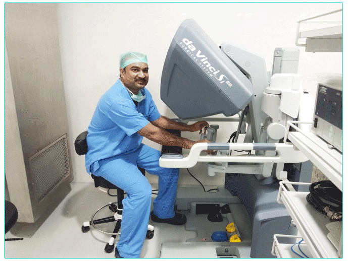
Hydatid disease is caused by infection with a small tapeworm parasite called Echinococcus granulosus.
People become infected by ingesting (eating) eggs of the parasite, usually when there is hand-to-mouth transfer of eggs in dog feaces.
The parasites form slowly enlarging fluid-filled cysts which may become very large. Cysts occur most commonly in the liver or lungs, but may occur in any organ, including the heart, brain and bone
Signs and symptoms
➠ Usually asymptomatic
➠ May present with abdominal pain,dyspepsia,fever with chills,jaundice etc.
➠ Usually presents with right upper quadrant mass and tenderness
Diagnosis
Diagnosed by x-ray, ultrasound, CT or MRI scans and may sometimes be confirmed by a blood test. Occasionally, microscopic examination of the cyst fluid is required.
Treatment
➠ Drug therapy – Albendazole and Mebendazole are used
➠ PAIR technique-Puncture,aspiration,Injection and reaspiration
➠ Endoscopic methods
➠ Surgical(open/lap)
Laparoscopic
➠ Done under general anesthesia
➠ A Special instrument has been developed for the removal of hydatid cyst with laparoscope called the PERFORATOR-GRINDER-ASPIRATOR APPARATUS
➠ This instrument penetrates the cyst,grinds the particulate matter and sucks it all out
➠ This technique is safe and simple
➠ The advantage is that it prevents intraperitoneal spillage of cyst contents.
The appendix is a small, tube-shaped pouch attached to large intestine. It’s located in the lower right side of the abdomen.
An appendectomy is the surgical removal of the appendix. It’s a common emergency surgery that’s performed to treat appendicitis, an inflammatory condition of the appendix.
Indications
➠ Acute appendicitis
➠ Perforated appendicitis
➠ Appendicular mass
➠ Appendicular abscess
➠ Chronic appendicitis
Symptoms
➠ Pain abdomen(mid and lower right abdomen)
➠ Nausea/vomiting
➠ Fever
➠ Loss of appetite
➠ Constipation or diarrhoea with gas
Treatment
Open
Done under general anesthesia.During an open appendectomy, a surgeon makes 2-3 inch incision in the lower right side of the abdomen,pulls the appendix through the incision,base is tied off and appendix is removed and the wound is closed with stiches.
Laparoscopic
Reduces post operative pain and lowers wound infection rate, return to work quicker and is also used as a diagnostic tool.
Cystogastrostomy is a surgery to create an opening between a pancreatic pseudocyst and the stomach when the cyst is in a suitable position to be drained into the stomach. This conserves pancreatic juices that would otherwise be lost.
A pancreatic pseudocyst is a circumscribed collection of fluid rich in pancreatic enzymes, blood, and necrotic tissue, Pancreatic pseudocysts are usually complications of Pancreatitis
Indications
Symptoms of this include abdominal bloating, difficulty eating and digesting food, and constant pain or deep ache in the abdomen. A lump can be felt in the middle or left upper abdomen if a pseudocyst is present.
To further diagnose a pancreatic pseudocyst an abdominal CT scan, MRI or Ultrasound can be used. Emergency surgery may need to be performed if there is a rupture of the pseudocyst.
Treatment
➠ Surgical cystogastrostomy
Surgical repair is carried out through an incision in the abdomen. After locating the pseudocyst, it is attached to the wall of the stomach and the cystogastrostomy is created.
➠ Endoscopic cystogastrostomy
. A large bore needle is used to access the identified pseudocyst, creating a fistula between the cystic cavity and either the stomach or the duodenum. Plastic stents may be placed to facilitate drainage from the pseudocyst.
➠ Laparoscopic cystogastrostomy
Laparoscopically, by a 3 port approach, gastrostomy is done on the anterior wall of the stomach.The pseudocyst is identified and accessed using laparoscopic techniques. Once the pseudocyst cavity is located, it is entered and aspirated, and an opening is created into the stomach for drainage. Laparoscopic drainage may result in better cosmetic appearance and decreased pain following surgery
What is fundoplication?
Fundoplication is a surgical procedure used to treat GERD ,Barrets oesophagus and symptomatic hiatus hernias in which the upper portion of the stomach is wrapped around the lower end of the esophagus and sutured in place as a treatment for the reflux of stomach content.This surgery strengthens the valve between the esophagus and stomach (lower esophageal sphincter).
Fundoplication surgical therapy
➠ Addresses the functional nature of GERD
➠ Restores anti-reflux barrier,strengthens oesophageal peristalsis,speeds gastric emptying,and improves gastric clearance
➠ Curative in 85-93% of patients
➠ Research of post-operative fundoplication patients have supported good long term results with low morbidity and mortality.
Open vs Laparoscopic
Open
➠ Incision of roughly 20 -25 cms in the abdomen
➠ Hospital stay – several days
➠ Recovery time – 4 – 6 weeks
➠ Indicated in patients who had multiple abdominal surgeries
Laparoscopic
➠ Takes about 1.5 to 2 hrs and is carried out under general anesthesia
➠ Minimally invasive technique producing five 0.5 -1cm incisions
➠ Hospital stay – 1 -2 days
➠ Recovery time – 2-3 weeks
Advantages of Laparoscopic Fundoplication
➠ Lesser blood loss
➠ Lesser rate of infections
➠ Lesser complications
➠ Minimal pain
➠ Faster recovery
Laparoscopic Hemicolectomy
A hemicolectomy is a surgical procedure that involves removing a segment of the colon. It is typically performed to treat colon cancer or a bowel disease, such as crohns disease or severe diverticulitis.
During this surgery, the damaged section of the colon is removed and the healthy parts of the colon are reattached.
Types
A hemicolectomy may involve removing a portion of the colon on the right or left side, depending on the location of the cancer or diseased bowel.
If a right hemicolectomy is done, the ascending colon is removed. The transverse colon is then attached to the small intestine.
During a left hemicolectomy, the descending colon, the section of the colon attached to the rectum, is removed. The transverse colon is then attached to the rectum.
Surgical procedures
Open hemicolectomy Done under general anesthesia.. Open surgery involves making a longer incision in the abdomen to access colon. Surgeon uses surgical tools to free the colon from the surrounding tissue and cuts out a portion of the colon.
Laparoscopic hemicolectomy Done under general anesthesia. Done by making few small incisions in the abdomen
After making the incisions, surgeon will remove the affected part of the colon and will also remove any parts of the intestines directly connected to the part of the colon being removed, any lymph nodes and blood vessels that are connected to the colon are also removed.
Once the affected part of your colon is removed, surgeon reconnects the rest of the colon. If ascending colon is removed, colon is attached to the end of small intestine. If descending colon is removed, rest of the colon is attached to the rectum. This rejoining is known as anastomosis.
Laparoscopic hemicolectomy is a safe option for cancers of the colon. It is associated with a shorter hospital stay and earlier resumption of a normal diet.
Surgical procedure to remove uterine fibroids,leaves the uterus intact
Fibriods are non cancerous growths that appear in the uterus usually during child bearing years
Removal is necessary when the fibroid causes pain,abnormal bleeding or interferes with reproduction
Management
➠ Observation
➠ Medical therapy-GnRH agonists
➠ Hysterectomy
➠ Uterine artery embolization
➠ High intensity focused ultrasound ablation
Surgical approach remains an important choice for women who wants to preserve uterus
Surgical
Abdominal myomectomy
Done under general anesthesia.Incision is made through skin on lower abdomen and fibroids are removed from wall of uterus.Uterine muscle is then sewn back together using several layers of stitches.
Hysteroscopic Myomectomy
Out patient surgical procedure.Scope is placed through cervix into the uterine cavity.Fluid is introduced to lift apart the walls.Instruments passed through hysteroscope are usedto shave off fibroids.Mainly done for submucosal fibroids.
Laparoscopic myomectomy
Done under general anesthesia
Safe technique with extremely low failure rate and good results in terms of outcome of pregnanacy.
Four small incisions given in lower abdomen .Laparoscope is placed through incision which allows to see ovaries, fallopian tubes and uterus. Long instruments inserted through other incisions are used to remove fibroids. Uterine muscle is sewn back together and skin incisions are closed.
Reduces hospitilisation stay ,post op pain,blood loss and recovery compared to open surgery.
Splenectomy is surgical removal of spleen
Spleen is a blood filled organ located in the upper left abdominal cavity.It is a storage organ for red blood cells and contains many specialized white blood cells called macrophages which act to filter blood.
Spleen plays a role in immunity against bacterial infections.
Indications
➠ Trauma
➠ Blood disorders like haemolytic anemia
➠ Enlarged spleen
➠ Benign tumors of spleen
➠ Autoimmune diseases
➠ Splenic cysts
➠ Leukemia or lymphoma
➠ Genetic conditions-herediatry spherocytosis and thalassemia
Types of Splenectomy
Open
The patient is administered general anesthesia.Surgeon makes an incision across middle or left side of the abdomen,muscles and tissue moved aside to reveal the spleen and is removed.Mainly done in trauma and hematological disesases.
Laparoscopic
Done under general anesthesia.Surgeon makes four small incisions in the abdomen,inserts a tube with a tiny video camera through one of the incisions.Surgeon watches video images on a monitor and removes the spleen with special surgical tools and incisions are closed.
Advantages
➠ Less postoperative pain
➠ Shorter hospital stay
➠ Faster return to a regular, solid food diet
➠ Quicker return to normal activities
➠ Better cosmetic results
➠ Fewer incisional hernias
Hysterectomy is the surgical removal of the uterus, it may also involve removal of the cervix,fallopian tubes,ovaries and other surrounding structures
Indications
➠ Fibroids,Adenomysis,Endometroisis
➠ Dysfunctional uterine bleeding ,Cervical cancer
➠ Uterine prolapse,Uterine cancer,Ovarian cancer
Types of Hysterectomy
Total hysterectomy- It is the surgical removal of the uterus and cervix
Partial hysterectomy- In this the uterus is removed ,but cervix is not removed
Radical hysterectomy- It is the removal of uterus,cervix,ovaries,structures that support the uterus. Done to treat endometriosis and cancers of uterus,cervix,ovaries.
Types According to Route
Vaginal- In this the uterus and cervix are removed through an incision in the top of vagina .Can be done to remove small uterine fibroids and when the uterus is of normal size
Abdominal- In this the incision is made in the abdomen horizontally or vertically.Recommended when uterus is very large,fibroids larger then 20cms,cancers and endometriosis.
Laparoscopic- It is the preferred treatment
Total laproscopic hysterectomyIs done by inserting a laparoscope and surgical instruments through several small incisions in the abdomen.Uterus and cervix are removed through one of the incisions Done to remove uterine fibroids which are small to moderate in size.
Laparoscopic supracervical hysterectomyIn this small insisions are made in the abdomen,uterus is removed but the cervix is left intact
Laparoscopic assisted vaginal hysterectomyIn this the surgical instruments are inserted through a vaginal incision and one or small abdominal incisions and the Uterus is removed through the vagina.
➢ Laparoscopic Cholecystectomy
➢ Laparoscopic Du Perforation Repair
➢ Laparoscopic Bowel Anastomosis
➢ Laparoscopic Pyeloplasty
➢ Laparoscopic Inguinal Hernia Repair
➢ Laparoscopic Ventral Hernia Repair
➢ Total Laparoscopic Hysterectomy
➢ MIPH
➢ Laparoscopic Endometriotic cyst, Ovarian Cyst, PCOD Drilling, Tubal Patency,Tubectomy, Ectopic Pregnancy

